Entering the new era of evidence based medicine, one would require a great deal of information about the functional and anatomical functions in the body before pinning down a diagnosis. For instance: if the differential diagnosis contains myocardial infarction (heart attack) then we must test for all functional aspects of a heart that may show signs of compromised muscular heart activity.
There would be two aspects of the doctor’s differential: patient’s condition and objective physiological or anatomical evidence. Patient’s condition may include: chest pain, sweating, shortness of breath, risk factors and history et cetera, whereas the physiological aspect would require us to check many things:
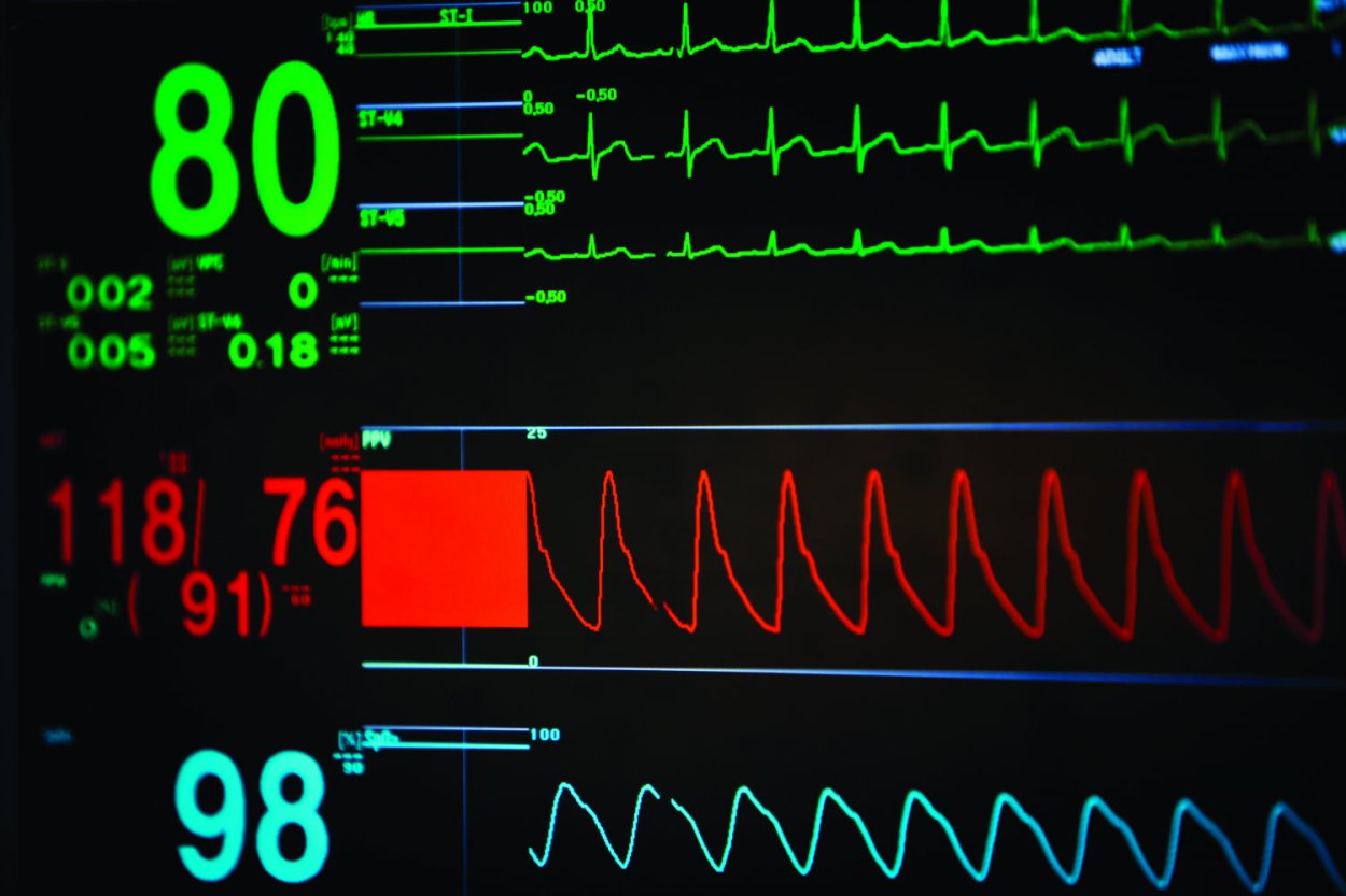
- The electrical activity of the heart that is required for the muscular pump’s contractions
- Checking if the heart is beating at a normal speed (Heart rate)
- Any chemicals specific to the heart that can be found in the blood to indicate that your heart is contracting fast and needs extra help to do so (Troponin levels in blood)
- Anatomical size of the heart and aorta (visualized using imaging)
These 4 things require some analysis and extraction that can be done using engineering tools. This is where biomedical engineering comes in. Using the inspiration and knowledge from medicine, we can construct tools to help us observe the exciting things going inside the body. In this example, engineering will help in quantifying and visualizing the 4 factors which can in-turn provide objective evidence for a diagnosis.
The primary aspect of the doctor’s differential that I will be exploring today is the electrical activity of the heart! As much as I would love to continue to rant about the heart and how it functions, for the sake of this post, I will limit that to the simplest possible explanation of just the electrical activity. In simple words: heart is a magnificent muscle pump. It contracts and it needs a signal to contract – that signal is an electrical impulse that magically generates from two places. For the sake of simplicity, let’s call them: island 1 (SA node) and island 2 (AV node).
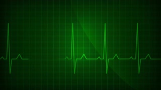
1- SA Node firing 2- AV Node firing
These nodes constantly fire (as shown in the images above) and are primarily the amazing things that make our heartbeat. When there is a current through island 1, then our small upper parts of the heart contract and when there is a current through island 2, it makes our two lower and larger chambers contract which then take the bloody awesomeness to other parts of the body.
In our case, if the heart isn’t functioning properly, there might be something wrong with this activity right? Either the current will be high or low. If its high, it could possibly mean that our body is trying super hard to get through something and is pushing signals harder, and if it’s low – these islands are probably having some internal technical difficulties.
So, the question is – how do we test this? Being a health enthusiast who would like to know the real time functionality of the heart, I’d wanna see some problem solving put into this. So let’s see how I would crack the code and find out what is going on inside the body.
To start off, lets walk through what we know from both worlds of engineering and medicine:
Medicine – Source of the problem Statement:
- What am I trying to do? Visualize the heart’s activity
- What do I know about the heart? Heart activity can be understood and interpreted by its electrical activity as the contractions correspond to the electrical signals. This tells us that the electrical activity and heart contractions are coupled, or in other words – directly proportional.
Engineering – Tool kit and problem solving algorithm:
- Some material that can respond to an electrical excitation, something that conducts electricity. Aha! Some electrodes will do
- But my electrodes can pick up so many other signals from the body –
Some extra components to isolate only the desired signal. - If I’m gonna use an electrically active material, I should have some safety net for the patient so they don’t get shocked: a safety circuit and some specific components for one way current flow
- Manufacturing awesomeness and screens to show the activity in real time
Now that we have all our blocks, lets get building!
Signal Detection

- Use electrodes to pick up the signal. As the electrical activity in the body is driven by ionic concentrations, we need to pick up those electrical changes (electrochemistry, boooo!).
- But, the voltage differences are so small that our electrodes can’t even pick it up until its amplified. To do that, we initiate a redox reaction in a gel electrolyte that corresponds to the body’s ionic changes.
- Body’s electrochemical activity –> electrolyte changes –> electrode excitation
Safety
- Add something to make sure that current flows in one direction. Also use something to wirelessly transfer the current to leave no room for electrons to **excitedly**
travel in the opposite direction. - We typically use a bridge for resistors and diodes along with isolation amplifier for these things.
Amplifying and Filtering
- Now we amplify the signal even more so that we can enhance its features and analyze it. In this step we need to find out the combination of resistors, amplifiers and capacitors that we want to use for the amplifying. As I wont be teaching amplifying circuits for a while, I’ll just say assume a black box that amplifies our signal by a magnitude of 50.
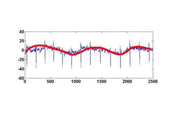
- Next would be filtering. For ECG machines, we usually place a filtering range after 0.5 Hz. This accounts for any low frequency noise in the ECG. For example, electrical activity associated with breathing breathing. Our breathing rate is approximately 20 per minute = 20/60 = 0.3 Hz. This will get cut off so that we can isolate the heart activity. If your ever moved during an ECG, you would probably see a low frequency noise (emphasized with red marker) like the one shown below.

Analyzing
- Now analyzing is the real fun part. I can calculate many things: heart rate, length of heart chamber contractions and many more things. For this part, we can connect our circuit above to a computer using a microprocessor, then after programming the microprocessor, the desired output for the heart activity can be displayed.
- I once did a project for just displaying the heart rate where I detected the peaks using Labview and then counted number of peaks in a minute to give me the heart rate. Additional information can easily be obtained by just doing time/ frequency analysis.
- And that is the starting point for an ECG machine. Once we have obtained this, we can expand it for printing, taking in a name input and even adding an artificial intelligence analysis tool for computer aided analysis of the ECG obtained.
A physiology expert can spit out how a signal comes in, fires an action potential, releases calcium ions followed by a lot of other steps ……… and then eventually a muscular movement, but as we saw today, understanding this relation can be further expanded into achieving an effective measuring tool. The question is, how do we bring this together for other applications? How do the health practitioners know what techniques they can use to visualize desired functional / anatomical features? Or, how do they even know that an engineering tool can be used to solve a problem if they don’t bring the problem to engineers cause it may seem to be too “medical” for them to inherit.
I believe the extraction of the electrical activity of the heart is a prime example of showing how this can be done. Once an engineering mind inherits the depth of physiological relations and finds inspiration from a medical practice, healthcare problems can be solved with creativity and this can yield great solutions at the intersection of medicine and engineering. In my future posts, I will further analyze problems in medicine that were resolved in the past using a strong problem solving mindset and how it transcended to the modern day – evidence based medicine – as we know it today. After this, I will further speak about newer problems that we can possibly explore to create a newer and even better healthcare practice.
